

Our Latest Research
Welcome to our research and publications page. Here, you'll find our latest research findings in the field of cancer biology, with a specific focus on mechanisms of metastatic initiation, dissemination, and outgrowth in pancreatic cancer. Our team is dedicated to understanding the underlying mechanisms of this disease and using that knowledge to develop new treatments and therapies to improve patient outcomes.
GSTT1 promotes stemness and FGFR inhibitor sensitivity in pancreatic cancer through regulation of CD133 (PROM1)

Pancreatic ductal adenocarcinoma (PDA) is among the deadliest malignancies, driven by metastatic progression and profound cellular heterogeneity. We previously identified glutathione S-transferase theta 1 (GSTT1) as a regulator of a slow-cycling, highly metastatic tumor cell population, suggesting that GSTT1High cells may possess stem-like properties. Here, we define the functional and molecular features of this subpopulation in metastatic PDA. Using a mCherry-tagged Gstt1 reporter system in metastatic murine PDAC cells, we enriched for Gstt1High cells and observed increased tumor sphere formation, accompanied by upregulation of stemness-associated genes including PROM1 (CD133) and activation of Wnt and FGF signaling pathways. In human PDA models, CD133HighGSTT1High cells exhibited enhanced tumor sphere initiation and expansion compared to other populations, defining a maximal stem-like state. Notably, sensitivity to FGFR inhibitors was observed only under tumor sphere conditions, highlighting a context-dependent therapeutic vulnerability. Mechanistically, FGFR3 expression correlated with GSTT1 and CD133 levels, and FGF signaling was required to sustain this state. GSTT1 knockdown reduced CD133 protein levels, impaired tumor sphere formation, and altered sensitivity to FGFR inhibition. These findings were largely recapitulated in patient-derived PDA organoids, where GSTT1 and PROM1 co-expression predicted increased tumor sphere formation and enhanced response to the multi-kinase inhibitor Nintedanib. Together, these results identify a GSTT1HighCD133High stem-like subpopulation in metastatic PDA and identify an FGFR-dependent signaling axis that sustains this state, representing a potential therapeutic vulnerability.
KRAS on Empty: Lipid Oxidation Blockade Reveals a Metabolic Achilles’ Heel in Pancreatic Cancer

Pancreatic ductal adenocarcinoma (PDAC) is the third leading cause of cancer death in the United States, driven by its aggressive biology and high metastatic incidence at diagnosis. With a 5-year survival rate of just 8%, PDAC remains one of the most lethal cancers. Mutant KRAS, present in more than 90% of cases, serves as a key driver of tumorigenesis and metabolic reprogramming. In this issue of Cancer Research, Thakur and colleagues uncover a novel metabolic adaptation that PDAC cells use to survive therapeutic stress. Their integrated metabolomic and lipidomic analyses show that ERK inhibition—targeting a key KRAS pathway effector—not only disrupts glycolysis and glutamine metabolism but also triggers a compensatory increase in fatty acid oxidation (FAO). This shift occurs through lipophagy, a lysosome-mediated lipid degradation process, rather than cytosolic lipolysis. Mechanistically, ERK inhibition promotes the nuclear translocation of TFEB, which drives the upregulation of FAO and lipophagy genes. This metabolic reprogramming enables PDAC cells to survive KRAS pathway blockade. Importantly, cotargeting FAO alongside ERK or KRAS inhibitors elicits a potent synergistic antitumor effect in vivo. This dual-target strategy holds promise for overcoming PDAC resistance to KRAS-targeted therapies, laying the groundwork for novel combination treatments.
The role of the immune tumor microenvironment in shaping metastatic dissemination, dormancy, and outgrowth
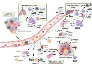
This review focuses on the tumor microenvironment (TME) is a dynamic and complex ecosystem composed of cancer cells and diverse non-malignant cell types, including immune cells, fibroblasts, and endothelial cells. Once viewed as passive bystanders, these host cells are now recognized as active participants in tumor progression, especially during metastasis. The TME varies by organ, cancer type, and disease stage, and shapes the trajectory of cancer progression. Among the immune cells in the TME, macrophages, neutrophils, and T cells play especially crucial and context-dependent roles - either promoting or inhibiting metastatic spread depending on the tumor stage, immune cell phenotypic states, and interactions. In this review we focus on the multifaceted contributions of these key immune populations across the major stages of the metastatic cascade: initiation, survival in the circulation, dissemination, dormancy, and reactivation. These insights highlight the heterogeneity of the metastatic immune microenvironment and underscore the therapeutic potential of targeting macrophages, neutrophils, and T cells to combat metastatic disease.
Identifying Unique Adaptations of Metastatic Pancreatic Cancer Cells

In this seminal manuscript, we describe a loss-of-function shRNA targeted screen in metastatic-derived cells, and identified the glutathione S-transferase Gstt1 as uniquely required for metastasis and dispensable for primary tumor growth. Within metastatic lesions, Gstt1 contributes to both intratumoral and intertumoral heterogeneity through regulation of proliferation, EMT and extracellular matrix deposition through glutathionylation of intracellular Fibronectin in a subpopulation of Gstt1high metastatic cells. These studies will significantly contribute to our understanding of unique vulnerabilities of metastatic pancreatic cancer and provide a possible therapeutic window for the treatment of a subset of patients with metastatic disease.
Understanding the Role of Gstt1 in Shaping the Metastatic Tumor Microenvironment
Here we are investigating the role of the tumor microenvironment in regulating the dissemination and outgrowth of a subset of pancreatic cancer cells, using lineage tracing and advanced imaging techniques to visualize the complex interactions between cancer cells and their surroundings. We hope to identify immune factors that promote or inhibit cancer cell spread and ultimately growth. This work is funded by an NIH-NCI R00 Pathway to Independence Award.


Identifying Metastasis-Initiating Cells in Pancreatic Cancer


This project uses lineage tracing techniques to identify whether a rare population of slow-cycling cells present in the primary tumor, preferentially disseminate and seed metastases in pancreatic cancer. We will identify and characterize key molecular pathways that contribute to the dissemination and switch to a proliferative state. This work is possible due to a Career Development Award in Honor of John Robert Lewis, funded by the AACR and the Lustgarten Foundation for Pancreatic Cancer Research.
Gstt1 Mediates Stemness and Sensivity to FGFR Inhibitors
A small, stem-like cell population drives the spread and treatment resistance of pancreatic cancer. Marked by high GSTT1 and CD133, these cells fuel tumor growth—but they may be vulnerable to targeted FGFR therapies. Our work points to new ways to tackle the deadliest forms of pancreatic cancer.
This work is funded by an NIH-NCI R00 Pathway to Independence Award.